

Unveiling the Microscopic World A Journey Through the Lens of a Microscope

Unveiling the Microscopic World A Journey Through the Lens of a Microscope
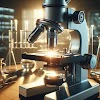
A microscope is a scientific instrument designed to magnify and observe objects that are too small to be seen by the naked eye. It works by exercising lenses or a combination of lenses to enlarge the details of bitsy structures, allowing for a near examination of natural, geological, or material samples.
Microscopes have been vital in advancing colorful fields of wisdom, enabling experimenters, scientists, and scholars to explore the complications of bitsy worlds. They come in colorful types, similar as optic microscopes that use visible light, electron microscopes that use electron shafts, and other technical forms, each acclimatized to specific operations.
The capability to claw into the bitsy realm has revolutionized scientific understanding, contributing significantly to advancements in drug, biology, chemistry, and accoutrements wisdom.
HISTORY OF MICROSCOPE : The history of microscopes is a fascinating trip that spans centuries, marked by the grim pursuit of understanding the bitsy world. Then is a brief overview of crucial mileposts
Early compliances( Age- 16th Century) : The conception of exaggeration dates back to ancient times, with early scholars using simple lenses made of glass or demitasse to observe small objects. still, the first recognizable microscope didn't crop until the late 16th century.
Invention of the emulsion Microscope( Late 16th Century) Dutch spectacle makers Hans Lippershey and Zacharias Janssen are frequently credited with creating the first emulsion microscope around 1590. It featured a convex objective lens and a hollow eyepiece, furnishing a significant enhancement in exaggeration.
Microorganisms Discovery( 17th Century) The microscope came necessary in revealing the actuality of microorganisms. In themid-17th century, Antonie van Leeuwenhoek, a Dutch scientist, perfected single- lens microscopes and observed bitsy organisms, laying the foundation for microbiology.
Advancements and advances( 18th Century) Microscopy saw farther advancements in the 18th century, with scientists like Joseph Jackson Lister introducing achromatic lenses, reducing color deformation in images. These advancements enhanced the clarity and quality of bitsy compliances.
The Rise of emulsion Microscopes( 19th Century) The 19th century witnessed significant developments in microscope design. benefactions by scientists like Carl Zeiss and Ernst Abbe led to the creation of high- quality emulsion microscopes, perfecting resolution and enabling detailed examination of natural samples.
Electron Microscope( 20th Century) The invention of the electron microscope in the 1930s marked a revolutionary vault in microscopy. Developed by Max Knoll and Ernst Ruska, electron microscopes use electron shafts rather of light, allowing for much advanced exaggeration and resolution.
Scanning Tunneling Microscope( 1981) The late 20th century saw the development of innovative microscopy ways, similar as the scanning tunneling microscope( STM), which enabled experimenters to fantasize individual tittles on shells.
Advancements in Modern Microscopy( 21st Century) moment, microscopy continues to evolve with slice- edge technologies likesuper-resolution microscopy, furnishing unknown clarity and perfection in imaging. ways like confocal microscopy and luminescence microscopy have come standard tools in colorful scientific disciplines.
The history of microscopes reflects a nonstop hunt for perfecting our capability to explore the bitsy realm, transubstantiating the way we perceive and understand the complications of life and matter at the lowest scales.
pan face="Söhne, ui-sans-serif, system-ui, -apple-system, Segoe UI, Roboto, Ubuntu, Cantarell, Noto Sans, sans-serif, Helvetica Neue, Arial, Apple Color Emoji, Segoe UI Emoji, Segoe UI Symbol, Noto Color Emoji" style="color: #374151;">
TYPES OF MICROSCOPE
There are different types of microscopes, each tailored to specific needs and offering unique opportunities for observing microscopic objects. Here you will find an overview of some common types of microscopes:
optical microscope: Compound microscope: Uses multiple lenses to magnify samples, with separate objective and eyepiece lenses. Suitable for various biological and medical applications.
Stereo Microscope (Dissection Microscope): Provides a three-dimensional view of larger samples, making it ideal for dissections and other applications requiring low magnification.
Electron microscope:
Transmission Electron Microscope (TEM): Uses electron beams to penetrate ultra-thin samples, providing detailed, high-resolution images. It is suitable for studying the internal structures of cells and materials at the atomic level.Scanning electron microscope (SEM):
Scans the sample surface with a focused electron beam, creating detailed three-dimensional images. Useful for studying surface morphology. Scanning probe microscope
Atomic Force Microscope (AFM): Scans the surface of the sample with a sharp tip and measures the forces between the tip and the sample. It provides high-resolution images and is widely used in nanotechnology and materials science.
Fluorescence microscope: Use fluorescence to visualize specific structures in a sample.Phase contrast microscope:
Increases the contrast of transparent and unstained samples by exploiting the phase differences of light passing through different parts of the sample. Useful for live cell imaging.
Dark field microscope: Illuminates the sample with grazing light, making it appear bright against a dark background. Increases the contrast of transparent color fields.
atOptions = {
'key' : 'a1a78d020cff5964f3f65ca3af54430c',
'format' : 'iframe',
'height' : 90,
'width' : 728,
'params' : {}
};
document.write('
Polarization microscope:
Equipped with polarization filters to study the optical properties of minerals, crystals and other anisotropic materials.
Digital microscope: Uses digital technology to capture and display images. Often equipped with features such as image capture, measurement tools, and compatibility with computer systems.
high resolution microscope: Overcomes the diffraction limit of light and achieves higher resolutions than conventional optical microscopes. Techniques such as stimulated emission depletion (STED) and structured illumination microscopy (SIM) fall into this category.
This type of microscope meets a variety of scientific and industrial needs, allowing scientists and professionals to examine and analyze microscopic structures with varying levels of detail and specificity.
OPTICAL MICROSCOPE
Optical microscopes, also called light microscopes, use visible light to magnify and observe samples. They are widely used in various scientific, medical and educational environments. Optical microscopes have evolved over time to offer different configurations and features. Here is an explanation of the main components and types of optical microscopes:Components of optical microscopes:
LenS : The primary lens closest to the sample and responsible for magnifying and creating the output image.
Eyepiece (spectacle lens): The lens closest to the viewer's eye, which further magnifies the image produced by the lens.
Illuminator: A light source that provides illumination of the sample.In most cases this is an integrated light source that is placed under the stage.
Scene : Platform on which a specimen is placed for observation.
condenser: An optical element that focuses and concentrates light onto a sample, thereby increasing contrast and brightness.
Aperture: Adjustable aperture under the condenser that controls the amount of light reaching the sample.Types of optical microscopes:
compound microscope:Uses multiple lenses to magnify the specimen.
Ideal for observing thin sections of specimens, such as cells and tissues.
Commonly used in biological and medical research.
Stereo Microscope (Dissecting Microscope):
Provides a three-dimensional view of larger specimens.
Suited for tasks like dissection, examination of surfaces, and manipulation of larger objects.It has separate optical paths for each eyepiece, ensuring depth perception.
Phase contrast microscope: Increases the contrast of transparent and unstained samples.
Particularly useful for imaging living cells and observing internal cell structures.
Dark field microscope: Illuminates the sample with grazing light, creating a bright image against a dark background.
Increases the contrast of transparent samples and is often used in microbiology.Polarization microscope:
Equipped with polarization filters to study the optical properties of anisotropic materials such as minerals and crystals.
fluorescence microscope: Use fluorescence to visualize specific structures in a sample.
Requires the use of fluorescent dyes or proteins and is often used in molecular and cellular biology.
Digital microscope: Includes digital technology for image capture and display.
Offers features such as image capture, measurement tools and compatibility with computer systems.Light microscopes play a key role in scientific research, education and medical diagnosis, offering versatility and accessibility across a wide range of applications. Technological advances are constantly expanding their capabilities, making them indispensable tools in various fields.
ELECTRON MICROSCOPE
Electron microscopes are powerful imaging tools that use electron beams instead of light to magnify and see details at the atomic and molecular levels. Electron microscopes, developed in the early 20th century, revolutionized the field of microscopy, allowing scientists to examine the ultrafine structures of materials and biological samples with unprecedented clarity. Here is an explanation of the main components and types of electron microscopes:
Electron microscope elements:
Electron Source: Typically a tungsten filament or electron gun.
Electronic lenses (condenser and objective): Magnetic or electrostatic lenses that focus and control a beam of electrons.
Sample Chamber: An area in which a sample is placed for observation. It is stored under vacuum to prevent diffusion of electrons.Electromagnetic Coils: Used to control the direction and focus of the electron beam.
Detector: Captures the electrons transmitted through or scattered by the specimen, producing an image.
Types of Electron Microscopes: Transmission electron microscope (TEM): Send electrons through an ultra-thin sample.
Provides high-resolution 2D images of internal structures at the atomic and molecular levels.Widely used in biological, materials and nanotechnology research.
Utilizing an electron microscope for scanning purposes
Scans the sample surface with a focused electron beam.
Generates detailed 3D images of sample surface morphology.
Commonly used to study the topography and surface structures of materials.
Scanning Transmission Electron Microscope (STEM):
Combines the functionality of TEM and SEM.It scans a focused electron beam through a thin sample, producing high-resolution images with detailed compositional information.
Advantages of Electron Microscopes:
High magnification: Electron microscopes can achieve much higher magnifications than optical microscopes, allowing the observation of nanoscale structures.
High resolution: Due to the short wavelength of the electrons, electron microscopes offer much higher resolution than their optical counterparts, enabling detailed imaging of fine structures.
Versatile: Electron microscopes are versatile instruments suitable for a wide range of applications, from biological research to materials science.
Depth of Field: SEMs provide a greater depth of field than optical microscopes, allowing clear images of three-dimensional structures on the sample surface. Despite their powerful capabilities, electron microscopes also have limitations, such as:
B. the need for a vacuum environment and the possibility of damage to samples by electron beams. However, their ability to reveal complex details at the nanoscale has made them essential to advancing scientific knowledge across all disciplines.user
Provides high-resolution 2D images of internal structures at the atomic and molecular levels.Widely used in biological, materials and nanotechnology research. Scanning electron microscope (SEM)
Scans the sample surface with a focused electron beam.
Generates detailed 3D images of sample surface morphology.
Commonly used to study the topography and surface structures of materials.
Scanning Transmission Electron Microscope (STEM): Combines the functionality of TEM and SEM.It scans a focused electron beam through a thin sample, producing high-resolution images with detailed compositional information.
IFRAME SYNC
OPERATIONS OF MICROSCOPE
1. Sample preparation: Fixation: A biological sample is subjected to fixation to preserve its structure and prevent its decomposition. Common fixatives include formaldehyde and glutaraldehyde.Embedding: The fixed sample can be embedded in a solid medium such as paraffin or resin to provide support for thin film cutting.
Slicing: A microtome is used to cut thin sections of the sample, creating slices suitable for microscopy.
2. Dye: Dyes and Stains: Biological samples are often stained to increase contrast and highlight certain structures. Various stains and stains highlight cellular components such as nuclei, cytoplasm, and organelles.
3. Optical microscopy operations: Microscope configuration: The microscope is configured according to the type of observation required (brightfield, phase contrast, darkfield, etc.).
Light Control: The intensity and angle of the light are adjusted to optimize contrast and brightness.
Focus Adjustment: The focus of the microscope is precisely adjusted to produce a sharp image of the sample.Changing the magnification: By using different lenses, the magnification can be changed, thus enabling a detailed examination of different structures.
4. Fluorescence microscopy work: Fluorescence labeling: Samples are labeled with fluorescent dyes or proteins that emit light when exposed to specific wavelengths.
Filter selection: Fluorescence microscopes use special filters to selectively pass the emitted light and thus increase the contrast.
Fluorescence imaging: The resulting fluorescence is recorded and provides information about the location and distribution of the labeled structures.
5. Electron microscopic operations: Sample Coating: In the SEM, samples can be coated with a thin layer of metal to improve conductivity and image quality.
Vacuum Operation: Electron microscopes operate under vacuum to prevent electron scattering, which requires careful sample preparation.
Electron Beam Adjustment: The intensity, focus and scanning parameters of the electron beam are adjusted to obtain optimal images.
6.Image acquisition and analysis: Digital Imaging: Images are captured using digital cameras or sensors integrated into the microscope.
Analysis Software: Image analysis software is widely used to measure, quantify and analyze the properties of captured images.
7. Interpretation and publication of data: Interpretation: Researchers interpret observed data in the context of their research questions.
Publication: The results are documented and published in scientific journals, thereby contributing to the expansion of scientific knowledge.Typically, surgical microscopes in biological research involve a combination of sample preparation, imaging techniques, and data analysis that allow scientists to examine the intricate details of biological structures.
MEDICINE: In medicine, microscopes play a key role in diagnosis, research and treatment. The operation of microscopes in medicine involves several important steps in the preparation, observation and analysis of biological samples. Here is an overview of these operations:
1. Sampling: Biopsy: Tissue samples are taken from patients and examined under a microscope. This is common in cancer diagnosis and other pathological tests.Blood smears: Blood samples are examined for abnormalities in the structure and number of cells.
2. Sample preparation: Fixation: Tissue samples are fixed with chemicals to preserve cell structures and prevent tooth decay.
Thin tissue sections are made with a microtome for microscopic examination.
3.Coloring:Histochemistry: Various stains and dyes are used to highlight certain tissue components to help identify abnormal cells or structures.
Immunohistochemistry (IHC): Antibodies labeled with fluorescent or chromogenic markers help identify specific proteins in tissues.
4. Operations of optical microscopy: Microscope Configuration: Microscopes are configured for specific observation techniques such as bright field, phase contrast or fluorescence microscopy.
Focus Adjustment: The focus of the microscope is adjusted to obtain clear images of cell structures.
Magnification: Different levels of magnification are used for detailed examination of tissues and cells.
5. Fluorescence microscopy work: Fluorescence labeling: Certain biomolecules or structures are marked with fluorescent dyes for better visualization.
Filter Selection: Filters are used to isolate specific wavelengths, improve contrast, and highlight specific cellular components.
6. Electron microscopic operations: Ultrastructural Analysis. Electron microscopes provide a detailed look at the ultrastructure of cells and tissues, helping to identify subcellular abnormalities. Detailed
Imaging: Samples are prepared and coated as needed to ensure optimal electron beam interaction and imaging.
7. Image acquisition and analysis: Digital Imaging: Microscopic images are taken with digital cameras for documentation and analysis.
Analysis Software: Image analysis software is used to measure, quantify and analyze pathological features.
Remote Diagnosis: Microscope images can be transmitted electronically for remote consultation, facilitating collaboration between medical professionals.
medical microscopes contribute significantly to diagnostic accuracy, research progress and treatment planning, thereby improving patient care and outcomes.
x-large;">
In the field of materials science, microscopes play a key role in characterizing and analyzing the structure, composition and properties of various materials at the microscopic and nanoscopic levels. The operation of microscopes in materials science involves several important steps in the preparation, observation and analysis of material samples. Here is an overview of these operations:1. Sample preparation: Section: Materials are prepared as thin sections or slices for microscopic examination.
Polishing: Material surfaces are often polished to obtain smooth, reflective surfaces, resulting in better microscopic results.Changing the magnification: For detailed examination, different lenses are used to vary the magnification.
2. Scanning electron microscopy (SEM): Coating: Non-conductive materials can be coated with a thin layer of conductive material (e.g. gold) to prevent charging during imaging.High-resolution imaging: SEM provides detailed 3D images of material surfaces and enables nanoscale observation of surface topography and morphology.
3. Transmission Electron Microscopy (TEM) Operations: Ultrathin Sections: Materials are cut into ultrathin sections to allow electrons to pass through the sample.
High Resolution Imaging:TEM provides ultra-high resolution images of internal structures at the atomic and molecular levels.
4.Atomic force microscopy (AFM) operations: Cantilever Scanning: The AFM uses a pointed tip attached to a protrusion to scan the surface of a material and provide topographical information.
Surface Characterization: AFM is used to analyze surface properties, measure roughness, and study nanoscale material properties.
5. Spectroscopic techniques: Micro-Raman Spectroscopy: Combines microscopy with Raman spectroscopy to provide information about molecular vibrations in materials.
Energy-dispersive X-ray spectroscopy (EDS): Integrated into electron microscopes, EDS analyzes the elemental composition of materials.
6. Image acquisition and analysis: Digital imaging: For documentation and analysis, images are taken with digital cameras integrated into microscopes.
Analysis Software: Image analysis software is used to measure material properties, analyze structures, and perform quantitative analysis.
7. In situ microscopy: real-time observations: Microscopes are used to observe material reactions, phase transitions and mechanical properties in real time, providing valuable insights into the behavior of materials under various conditions.
8. Quality control microscopy: Defect Analysis: Microscopes are used for quality control and enable the identification and analysis of defects or anomalies in materials.
Microscopes in materials science contribute to advances in the development of new materials, understanding material properties and improving manufacturing processes. These processes are essential for researchers and engineers when characterizing and developing materials for specific applications.
FORENSIC SCIENCE Microscopes are indispensable tools in forensic science. They allow investigators to examine and analyze evidence, identify substances, and collect information critical to criminal investigations. The operation of microscopes in forensic science involves several phases of preparing, observing and analyzing evidence. Here is an overview of these operations:
1. Trace investigation: Hair and Fiber Analysis: Microscopes are used to examine the morphology, color, and characteristics of hair and fibers found at crime scenes or on suspects.
DNA analysis: Microscopic Analysis of DNA Samples: Microscopes are used to examine DNA samples during forensic DNA analysis and help identify genetic markers and assess sample quality.
3. Analysis of bloodstain patterns: Microscopic Examination of Bloodstains: Microscopes help forensic analysts examine bloodstains for patterns, thereby helping to reconstruct events at crime scenes.
4. Inspection of tools and firearms: Microscopy for Tool Mark Detection: Microscopes are used to analyze tool marks left at crime scenes or on evidence, helping to match tools to specific markings.
Case and cartridge analysis: Bullets and cartridge cases are examined using microscopes to identify clear traces of firearms.
5. Document verification: Ink Analysis: Microscopes facilitate analysis of ink composition and writing properties, helping to identify the source of written materials.
Paper Analysis: Microscopic examination involves analyzing paper fibers, watermarks, and other features to determine the authenticity and provenance of documents.
6.Drug analysis: Microscopic Examination of Drug Samples: Microscopes are used to examine illegal drugs and help identify substances, analyze their purity, and collect evidence in drug cases.
7. Soil analysis and geology: Microscopic Soil Examination: Microscopes help forensic geologists analyze soil samples and help link suspects or crime scenes based on soil properties.
8. Forensic Microbiology: Microscopic Examination of Microorganisms: In cases of bioterrorism or infectious diseases, microscopes facilitate the identification and analysis of microbiological evidence.
9. Trace metal analysis: Microscopic Examination of Trace Metals: Microscopes are used to analyze trace metal particles and link suspects to crime scenes or weapons.
10. Digital microscopy in digital forensics: Microscopic Analysis of Digital Evidence: Forensic experts use digital microscopes to analyze digital evidence, such as examining the microstructures of electronic devices, detecting tampering, or recovering deleted data.
11.Crime scene documentation: Microscopic Photography: Microscopes equipped with cameras are used to capture detailed images of microscopic evidence for documentation and presentation in court.
12. Comparative microscopy: Direct Comparisons: Microscopes allow direct comparisons between recognized and known samples, helping to identify similarities or differences.
Forensic microscopes play a key role in discovering evidence, solving crimes and providing important information in legal proceedings. The detailed observations made possible by microscopes contribute significantly to the investigative process, helping forensic scientists draw accurate conclusions and support their findings in court.
ASSIDUITY & QUALITY CONTROL
In industry and quality control, microscopes are important tools for examining materials, assessing product quality and ensuring compliance with strict standards. The operation of microscopes in industry and quality control involves several important steps to analyze materials and detect possible defects or irregularities. Here is an overview of these operations:
1. Control of raw materials: Microscopic Inspection: Microscopes are used to inspect raw materials for impurities, defects, or irregularities that could affect the quality of the final product.
2.Monitoring the production process: In-Process Inspection: Microscopes are used to monitor various stages of the manufacturing process and ensure that components are manufactured to required specifications.
3. Surface inspection: Surface Defect Detection: Microscopes, particularly scanning electron microscopes (SEMs) or optical microscopes, are used to examine surfaces for defects, cracks, or imperfections that could affect product quality.
4. Material analysis: Microstructure Analysis: Microscopes help analyze the microstructure of materials, providing information about their composition, particle size and overall quality.
Metallography: Microscopic examination of metal structures allows you to evaluate the integrity of materials and identify potential problems such as porosity or inclusions.
5. Quality assurance in electronics: Printed Circuit Board (PCB) Inspection: Microscopes are used to carefully inspect circuit boards for soldering defects, poor connections, or other electronic component problems.
6. Testing the coating and surface quality: Microscopic Analysis of Coatings: Microscopes are used to evaluate the uniformity and quality of coatings on surfaces and ensure that they meet specific thickness and coverage requirements.
7. Particle analysis: Contaminant Detection: Microscopes are used to identify and analyze particles or contaminants on surfaces or materials to ensure cleanliness standards are met.
8. Dimensional Analysis: Microscopic measurements: Microscopes equipped with calibrated scales or measuring instruments help assess the dimensional accuracy of components and ensure that they adhere to specified tolerances.
color: #374151; white-space-collapse: preserve;">
9.Error analysis: Microscopic Defect Investigation: Microscopes play a key role in investigating product defects by examining microscopic details of faulty components, identifying root causes, and initiating corrective actions.
10. Quality control of weld seams: Microscopic Inspection of Welds: Microscopes are used to inspect welds for defects such as porosity, incomplete fusion, or improper penetration to ensure structural integrity.
11. Inspection of fibers and fabrics: Fiber Analysis: Microscopes help examine textile fibers for quality, length and irregularities that can affect the strength and durability of the materials.
12. Pharmaceutical quality control: Microscopic Analysis of Pharmaceutical Products: Microscopes are used to examine the quality and properties of pharmaceutical products, ensuring uniformity, particle size and freedom from contamination.
The industrial and quality control microscopes contribute significantly to maintaining high standards, minimizing errors and ensuring the consistency and reliability of the products manufactured. The detailed observations provided by microscopes help industries meet quality requirements and deliver products that meet or exceed customer expectations.

.jpeg)


.png)
.png)
0 Comments